The Impact of AI and Computer Vision on Medical Diagnostics

For many years, diagnostic medicine functioned under a heavy load of doubt. Pathologists tediously reviewed glass slides on microscopes, frequently overlooking initial evidence of cancer due to exhaustion or visual confusion. Radiologists were blinded by the sheer number of medical images, and human mistakes became a constant risk. In country clinics, specialists were not available or were in short supply, causing life-saving diagnosis to be delayed. The chances of detecting microscopic tumors, faint fractures, or degenerative diseases in their earliest stages usually seemed like a needle in a haystack — too late, too problematic, or too dependent on the chance of getting the right picture at the right moment. The consequences were huge, and the room for mistakes was frightfully narrow. It was against this backdrop that computer vision came along.
Computer Vision is revolutionizing diagnostics in ways that have long been regarded as science fiction. From automating cancer detection in pathology slides to enhancing radiology workflows and bringing AI-powered image analysis to the forefront, this field has reshaped modern medicine. Computer Vision engineers have leveraged deep learning, convolutional neural networks (CNNs), and cutting-edge picture processing algorithms to build diagnostic applications that assist, hasten, and even outpace humans in terms of accuracy and efficiency in most cases.
In this article, we take a deep dive into the changing landscape of AI-assisted diagnostics and how it is impacting the way we deal with healthcare challenges.
Pathology: AI-Powered Microscopy and Slide Analysis
Automated Cancer Detection – One of pathology’s most significant breakthroughs has been employing deep learning to diagnose cancer in histopathology slides. Whole Slide Imaging (WSI) platforms with capabilities powered by ResNet, EfficientNet, and Vision Transformers (ViTs) have been trained with massive datasets and can accurately detect malignant regions in tissue samples. Software tools such as Google’s LYNA (Lymph Node Assistant) have demonstrated levels of accuracy in metastatic breast cancer diagnoses which are equivalent to those of experienced pathologists.
Digital Pathology and Image Enhancement – Traditional pathology involves cumbersome slide preparation and manual examination. Computer vision-enhanced applications like OpenSlide and QuPath digitize slides at high resolutions, and they can then be leveraged for AI analysis. Stain normalization (with CycleGANs) and feature extraction (with autoencoders) have been added to methodologies to improve the reliability of AI-powered evaluations.
Biomarker Detection and Quantification – In immunohistochemistry (IHC), VGG16 and InceptionNet deep neural networks can now quantify biomarker expression levels, enabling proper disease prognosis for breast and prostate cancer, for example. Paige.AI and PathAI have even gone a step further and designed AI algorithms that can classify tumors based on their probability of malignancy and provide crucial precognitive data for oncologists.
Real-Time Pathology with Edge AI – Edge AI platforms, including NVIDIA Jetson and Intel OpenVINO, enable real-time analysis of images in microscope-integrated platforms. These can help AI-assisted diagnostics to produce real-time results with zero cloud-based inference, opening doors to high-tech pathology even in rural settings.
Radiology: AI as a Second Opinion and Workflow Optimizer
Medical Image Segmentation and Annotation – Segmenting anatomical structures in X-rays, MRIs, and CT scans is a tedious process. AI algorithms using U-Net, DeepLabV3+, and Mask R-CNN have heightened segmentation accuracy to a considerable level and have aided radiologists in identifying regions of interest such as lung nodules, brain tumors, and fractures.
AI-Powered Chest X-Ray Analysis – Chest X-rays are one of the most prevalent radiographic exams, so bringing automation to this process was critical to reduce human errors. Models such as CheXNet, having been trained on millions of X-rays, have successfully diagnosed pneumonia, tuberculosis, COVID-19, and many such ailments. These algorithms use DenseNet architectures to provide high sensitivity in identifying pathologies that even a seasoned radiologist can miss.
MRI and CT scans – Traditional CT scans were notorious for the unchecked radiation exposure they forced patients to endure. Low-dose CTs have done away with that issue, but at the cost of compromised image resolution. Engineers have been trying to rectify this using Generative Adversarial Networks (GANs) and super-resolution techniques, the results of which can be seen in the deep learning-enhanced MRI images being generated by GE Healthcare’s AIR Recon DL.
Automated Fracture and Lesion Detection – Fractures and minor lesions have long been notoriously difficult to detect. YOLO (You Only Look Once) and RetinaNet-powered AI algorithms have been integrated with PACS (Picture Archiving and Communication Systems) for automatically detecting and highlighting suspected abnormalities in X-rays and CT scans, with a reduced chance of errors in diagnoses.
Radiomics and predictive analytics – Radiomics involves quantitative feature extraction from medical images for identifying disease signatures. AI radiomic models use algorithms such as XGBoost and LightGBM to analyze imaging biomarkers for disease progression, therapeutic response, and prognosis prediction in a patient.
Medical Imaging: Beyond Traditional Diagnostics
Ultrasound Automation and AI-Guided Scanning – Ultrasound imaging has seen a significant boost from applications of AI, particularly in automated cardiac and fetal assessments. AI-enabled ultrasound guidance platforms, including Butterfly Network’s AI-guided probes, leverage computer vision to allow practitioners to acquire optimal images with reduced operator reliance and increased diagnostic consistency.
Ophthalmology and Retinal Image Analysis – AI has turned out to be extremely efficient in ophthalmology, specifically detecting age-related macular degeneration (AMD) and diabetic retinopathy. Google DeepMind created an AI model utilizing VGG19 and InceptionV4 architectures that can assess retinal fundus images as efficiently as trained eye-care experts.
Endoscopy and Colonoscopy – Ask any oncologist and they would say the very thought of trying to detect polyps and early-stage colorectal cancer gave them nightmares even a few years ago. Not anymore though. AI-powered endoscopy systems such as Medtronic’s GI Genius now use deep learning to mark suspicious regions of the gastrointestinal tract in real-time, raising early detection rates significantly.
Dermatology and early Skin Cancer Detection – AI-enabled skin cancer diagnosing software have integrated deep neural networks with architectures like EfficientNet and MobileNet, that have been trained on massive datasets of thermostatic photos. The result is that, applications such as SkinVision can now automatically classify a lesion as malignant or benign and enable early intervention.
3D Reconstruction for Surgery and Planning – AI-powered 3D medical image reconstruction helps surgeons plan complex surgical operations. 3D Slicer and MONAI, for example, allow high-fidelity anatomical modeling with MRI or CT scan data, enhancing robotic and preoperative planning operations.
How Computer Vision is Changing Diagnostics for the Better
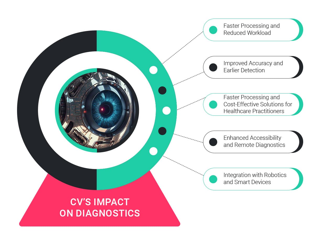
Improved Accuracy and Earlier Detection – AI-driven computer vision reduces the chances of missed diagnoses at a considerable level. By conducting in-depth analysis of massive amounts of imaging data, deep learning algorithms can detect minor abnormalities that even expert eyes can overlook, providing early intervention opportunities and enhancing patient care.
Faster Processing and Reduced Workload – Radiologists and pathologists have increasingly high workloads nowadays, but AI-guided diagnostics can ease this burden. Automated reporting and analysis save time, allowing for accelerated patient care with no loss in quality.
Cost-Effective Solutions for Healthcare Practitioners – AI-powered diagnostic platforms minimize repeat tests, errors and overall medical expenses to a bare minimum. AI platforms in cloud environments like IBM Watson Health and Microsoft Azure AI allow hospitals to introduce easily scalable diagnostics with little to no infrastructure investments in terms of hardware.
Enhanced Accessibility and Remote Diagnostics – AI-driven diagnostic platforms have made expert-level analysis accessible in regions with poor medical infrastructure. AI-powered cloud-accessible pathology analysis and mobile ultrasound machines have brought high-quality diagnostics to rural and disadvantaged communities.
Integration with Robotics and Smart Devices – Robot-assisted surgeries and AI-guided interventions have become widespread nowadays. AI-powered robotic platforms, such as the Da Vinci Surgical System, have reduced surgical errors through computer vision-guided operations and have improved patient rehabilitation efforts.
What the Future Holds
Computer vision has fundamentally altered pathology, radiology, and medical imaging operations at a grass-root level. From early cancer detection to robotic surgical guidance, AI-powered image analysis is redefining diagnostics. With deep learning, federated AI to safeguard patient information, and real-time edge processing, medical diagnostics in the future will become smarter, faster, and more accessible than ever before. Medical professionals and engineers together have become the pioneers of this revolution, developing machines that will go on to save lives with AI-powered precision medicine.
VE’s computer vision specialists are invaluable assets for every enterprise that is considering developing their own computer vision capabilities. Tell us what you need and our experts will get back to you right away.